Dolphin Slaughter

0
Here's an excerpt from an article on artificial intelligence, and includes dolphin intelligence in comparison with human.

Is Artificial Intelligence Possible?
By Tommy Connolly
By many of the physical methods of comparing intelligence, such as measuring the brain size to body size ratio, cetacean surpass non-human primates and even rival human beings. For example “dolphins have a cerebral cortex which is about 40% larger a human being. Their cortex is also stratified in much the same way as humans. The frontal lobe of dolphins is also developed to a level comparable to humans. In addition the parietal lobe of dolphins which "makes sense of the senses" is larger than the human parietal and frontal lobes combined. The similarities do not end there; most cetaceans have large and well-developed temporal lobes which contain sections equivalent to Broca's and Wernicke's areas in humans.”
Dolphins exhibit complex behaviours; they have a social hierarchy, they demonstrate the ability to learn complex tricks, when scavenging for food on the sea floor, some dolphins have been seen tearing off pieces of sponge and wrapping them around their "bottle nose" to prevent abrasions; illustrating yet another complex cognitive process thought to be limited to the great apes, they apparently communicate by emitting two very distinct kinds of acoustic signals, which we call whistles and clicks.
One example of their dissimilar brain structure and intelligence is their sleep technique. While most mammals and birds show signs of rapid REM (Rapid Eye Movement) sleep, reptiles and cold-blooded animals do not. REM sleep stimulates the brain regions used in learning and is often associated with dreaming. The fact that cold-blooded animals do not have REM sleep could be enough evidence to suggest that they are not conscious and therefore their brains can definitely be emulated. Furthermore, warm-blood creatures display signs of REM sleep, and thus dream and therefore must have some environmental awareness. However, dolphins sleep unihemispherically, they are “conscious” breathers, and if fall asleep they could drown. Evolution has solved this problem by letting one half of its brain sleep at a time. As dolphins utilise this technique, they lack REM sleep and therefore a high intelligence, perhaps consciousness, is possible that does not incorporate the transitional states mentioned earlier.
The evidence for animal consciousness is indirect. Intriguing, but more proof is required. However merely because we do not understand something does not mean that it is false - or not. Studying other animal minds is a useful comparative method and could even lead to the creation of artificial intelligence (that does not include irrelevant transitional states for an artificial entity), based on a model not as complex as our own. Still the central point being illustrated is how ignorant our understanding of the human brain, or any other brain is and how one day a concrete theory can change thanks to enlightening findings.

The Inhumanity of Japan's Dolphin Slaughter
This is a very graphic video I stumbled upon, on youtube.com, and disturbing as the footage was, I feel people need to know what's going on. Japan's food industry should be held in account for this barbaric practice. Dolphins have complex neural networks not too very different from humans and should not be treated in this inhumane way. Dolphin Slaughter, on youtube.com
Herding Dolphins into Bay

Dolphins are herded into a small bay by banging on pipes which interfere with the dolphin's sonar (echolocation, hearing), confusing and frightening them.
Dolphin Slaughter in Japan
Some dolphins are sorted and sold to aquariums while others are brutally stabbed and rounded up and dragged off to the slaughter house.
Dolphin Slaughter in Japan
Machettis are used to cut open throats of conscious dolphins as they lay helpless on the pavement...
Dolphin Slaughter in Japan
Blood pouring from this dolphin's throat...
Dolphin Slaughter in Japan
Dolphin going in shock and writhing in agony...
Dolphin Slaughter in Japan
School children who walk by in this video can see the dolphins agonizing in painful convulsions and slowly dying.
Dolphin Slaughter in Japan
Japan's indifference to the dolphin slaughter shows it has no respect for regulations regarding international waters, which harm both the ocean and creatures that live within them.
Dolphin Slaughter in Japan
Last year, I transferred an article to the web which was printed in the May 1911 issue of National Geographic, on the "Shore Whaling Industry by Roy Chapman Andrews, Assistant Curator of Mammals, American Museum of Natural History. A guide through the grisly slaughter of whales, an industry that flourished between the nineteenth and twentieth century.
Roy C. Andrews writes in 1911: "And what is to be the result of this wholesale slaughter? Inevitably the commercial extinction of the large whales, and that within a very few decades. In some localities this has already taken place and all the whales have been killed or driven from their feeding grounds."
Dolphins are Endangered
"Other dolphin species in different regions also need protection. The UN Environment Programme (UNEP) works with governments around the world to identify biodiversity-rich areas and designate specially protected areas for wildlife. For example, in the Caribbean and other regions, UNEP is supporting projects to protect the marine environment, making it safer for dolphins and whales.
As part of the global effort to protect the planetâs biodiversity, UNEP administers one of the world's largest conservation agreements-the Convention on International Trade in Endangered Species of Wild Fauna and Flora, known as CITES. Adopted in 1973, it became international law two years later.
More than 150 governments have ratified the treaty, which offers varying protection to more than 35,000 species of animals and plants, depending on their condition in the wild and the effect that international trade may have on them. CITES bans international commercial trade in species threatened with extinction, such as cheetahs, tigers, the great apes, many tortoises and birds of prey. It also protects other species, which are not threatened, but may be at serious risk unless international trade is strictly regulated."
- Dolphins as Endangered Species, United Nations Website

WHALES ARE DEVOTED TO THEIR CALVES
"All the large whales show great affection for their young, and the cows and calves will seldom leave each other when pursued by a ship. I remember at one time in Alaska, on board the steamship Tyce, Jr., we had sighted a female finback with a young one about 30 feet long beside her. They were not difficult to approach, and as the old whale rose to spout not five fathoms from the vessel's nose, the gunner fired, killing her almost instantly. The calf, although badly frightened, continued to swim in a circle about the ship, and finally, when its dead mother had been hoisted to the surface, the little fellow came alongside so close that I could have struck him with a stone. During the time that the carcass was being inflated and the gun reloaded, the calf was constantly within a few fathoms of the ship, swimming around and around, sometimes rubbing itself against the body of its dead mother. Finally a harpoon was sent crashing into its side, and it sank without a struggle."
- Roy Chapman Andrews, 1911

----------
Sample Letter to Japanese Officials, (seashepherd.org)
I am outraged by the annual brutal slaughter of dolphins and whales that takes place in Japan. The images of bloody red water clearly show the world that Japan has little respect for the state of the world’s oceans and for the conservation of the marine resources it claims to support.
Many scientific studies show that the oceans are in decline. We must take whatever actions are necessary to stop their over-exploitation and to protect the creatures that live in them. These dolphins do not belong to Japan. The status of the species of dolphins and whales that you kill are either endangered, threatened, or unknown. It is an unthinkable waste that they will likely end up as a meat product or deceptively sold as whale meat, polluted with toxic levels of mercury and cadmium, killing people that eat it. It is tragic and unacceptable that the remaining dolphins that are not killed will end up destined for death in an aquarium, water park, or "swim with dolphins" program.
In addition, the methods used to kill these animals are cruel. Corralling the dolphins into bays, then making them suffer a long and painful death by spears, hooks, and drowning is an inhumane way of fishing. This action is disgraceful and has caused much disappointment in the international community.
We demand that Japan permanently and immediately renounce and stop this slaughter. We will work diligently to bring this issue to international light until you have ceased your reprehensible violence.

Sincerely,

Read More »

Spirogyra

0
Recently, the instructors at Brunswick took myself and another student to the ponds and showed us certain common elements that are important to proper pond management and care. One of the important things I learned was about filament algae, and the threat it poses to fish.
Algae Pond Scum
Pond with filamentous algae, at Brunswick Community College
Enlarge to full 2816 x 2112 pixels resolution


Filamentous Algae
Filamentous algae, at Brunswick Community College
Enlarge to full 2816 x 2112 pixels resolution

Filamentous algae are microscopic algae that form colonies of “filaments” — hence the name. These algae are notorious for forming the large, pillow-like mats of algae that float on the surface of ponds. Common types found in Ohio include Spirogyra and Pithophora.
Disadvantages
As in the case with planktonic algae, high levels of nutrients can cause filamentous algae abundance to explode, especially in ponds lacking other aquatic plants, becoming so abundant that severe oxygen problems can result in the pre-dawn hours during July and August. Treating a severe filamentous algae problem in summer will almost certainly cause a fish kill. Ohio State University Extension Fact Sheet A-8-01, Winter and Summer Fish Kills in Ponds, provides insight into how these types of summer kills occur.
From Benefits and Disadvantages of Aquatic Plants in Ponds, Ohio State University Extension
Filamentous Algae

Filamentous Algae

Algae are primitive aquatic plants that differ from other plants in that they have no true stems, leaves or roots. They have a place in the overall food chain as they convert the energy of the sun into forms that can be used as a food source for other aquatic life. Algae also help to increase dissolved oxygen in water. Algae grow in both fresh and salt water systems. There are said to be over 20,000 different named species of green algae. Algae occur in three different basic forms. These are categorised as planktonic, filamentous and macrophytic.
Worldwide, there are over 400 different species of the genus Spirogyra. Spirogyra tends to show in ponds as a tangled pond scum. It is also called “water silk”, “silk weed” and “mermaid tresses”. On sunny days, the mats of spirogyra filaments usually float on the surface of the water. They are kept afloat by tiny bubbles of oxygen arising from photosynthesis. These algal mats then sink when the sun goes down and the process reverses as photosynthesis is reduced. As a result, the strands of Spirogyra consume oxygen for cellular respiration. Carbon dioxide is then produced as a waste product. Where there are thick algal mats present, large fluctuations in the dissolved carbon dioxide and oxygen levels in the water can occur. This can lead to rapid changes in the pH of the water that in turn can cause stress and even death to other organisms, eg fish, living in the water.
Blanket Weed and other Pond Algae


Spirogyra
Spirogyra - A Filament Algae, magnified at +/- 40x
Spirogyra

Spirogyra

Spirogyra

Spirogyra

Spirogyra

Spirogyra
A Desmid and Spirogyra algae found in the pond sample.

Cladoceran
A Cladoceran found among the algae sample.
Read More »

Vorticella - Protozoan Parasite

1
One of the protozoans I recently photographed, is Vorticella.
A few evenings ago I captured multiple videos with the same organism.. heavily populated slide with these flower-like contractile stalk organisms.
http://pirx.com/droplet/gallery/vorticella.html contains a clear photograph of this organism I compared with, among others on the web.

"Vorticella is a sessile, peritrich ciliate. Disturbed contracts and myoneme becomes spring-like. The stalk is not branched. Often forms clusters, but not colonies."

I saw two of the stalks interacting to some degree, it's pretty amazing. This was one of my favorites that shows their behavior..
http://www.youtube.com/watch?v=Gn_z5uCokDI They seem to be quite aggressive. These are a parasite in prawns and crawfish, but it seems there's disagreement among culturists / researchers, if its a "most common" or "least common" parasite.

"Hall (1979) found that Corthunia sp, Epistylis sp. and Vorticella sp. were the most common peritrichous ciliates in cultured prawns. Common sites of infestation are the body, eye stalk, antenna, uropods and egg masses. Thelohania, a microsporidian, has been reported in various species of marine shrimps but rarely in freshwater prawns. Areerat (1988) reported one case of microsporidia infection in the opaque muscular tissue of Macrobrachium."
From Diseases of the Freshwater Prawn, Aquatic Animal Health Research Institute

(Our Fundamentals of Aquaculture text, was likely the source I was citing from):
"Ectocommensals are comprised of a variety of protozoan species which live on and/or attach to the surface of the body and the gills of their host. Common parasitic genera associated with crawfish and prawns include Epistylis, Zoothamnium, Lagenophrys, Corthunia and Acineta. Less common genera are Vorticella, Vaginicola and Opercularia."

Anyway.. both agree they're parasitic on prawns and crawfish.

Vorticella spewing waste or another bi-product? black dots / fluid begin spewing from it's bud? (about half-way through video).
http://youtube.com/watch?v=VSzc-Cl8M4Q
Vorticella
Vorticella

Vorticella makes an interesting video subject.
Video #9, Video #8, Video #7, Video #6, Video #5, Video #4, Video #3, and others on youtube.com
Read More »

Fish Dissection

2
Preliminary Student Dissection of Fish
Head and Gills -- high magnification and some microscopy of gills, and various sections from gills to head.

This is for my term project in Aquaculture Practicum. I'm closely observing details as I go through the fish and then to study about certain features and processes in fish anatomy, and last create a model/s of those important parts of a fish.

Warning: Some of the images are graphic, while others are enlightening in understanding how a fish (as a machine) works.
Some of the less graphic images are as follows:
Fish Eye Lens
Lens of Eyes in a Fish.
Fish teeth
Staggered teeth growth on lower palette.
Fish Mouth
Fully expanded fish mouth.
Magnified fish gills
Magnification of a Fish Gill at about +/- 100x
Read More »

Teaching a man to fish...

0
Today I visited the local fish market for "fish heads" to do part of my fish dissection project. I want to investigate how the brain of a fish works and interconnected. They were very kind and supplied me with a bag full of fish heads, on the house. *smile*

I saw they had this neat bumper plate in the window.
teach a man to fish
Read More »

Yellow Perch

0
Yellow Perch Production
Modified in parts from class notes by Dr. Doug Holland, Aquaculture Program, Brunswick Community College

Yellow perch (Perca flavescens) is a relatively new species to aquaculture. This species is widely distributed, ranging from Nova Scotia to South Carolina along the Atlantic seaboard and throughout the Great Lakes region and as far west as the Dakotas and Nebraska. Commercially, it has been cultured in the upper Midwest, and in the Eastern US from Pennsylvania to South Carolina.

The best culture method for yellow perch is widely debated. The material presented here is based on the extensive experiences of staff at Brunswick Community College and yellow perch farmers in Brunswick County, North Carolina.

Broodstock Management and Egg Collection
Eggs may be collected from the wild, but as with all cultured aquatic species, preferred stock comes from domesticated broodstock. In North Carolina, one of the best places to collect wild eggs is from the Perquimmans River, a tributary of Albemarle Sound. The NC Division of Marine Fisheries manages the waters below the old bridge in downtown Hertford, and there is a healthy commercial fyke-net fishery for yellow perch in these waters. The perch are trapped in the nets during their annual spawning runs in February and March, and fertilized egg ribbons may be easily collected from the nets where the fish spawn. Egg ribbons may also be collected in submerged vegetation along the edges of the river.

Domesticated yellow perch broodfish may be held in ponds at densities up to about 500 lbs/acre. The fish should range from ¼ lb to 1 lb+ in size, and there should be a 50/50 mix of males and females. Good egg production generally requires an annual increase in body weight of at least 50%. Broodfish that are feed-trained will grow and reproduce well on pelleted feeds containing about 40% crude protein and 10% crude fat. Yellow perch generally prefer live food and use of a forage species such as fathead minnows in brood ponds, which will likely improve egg production and success with spawning.

Spawning
Spawning in ponds begins in mid-February. It is a good idea to encourage growth of submerged aquatic vegetation around the edges of brood ponds for the perch to use as a spawning substrate. If there no submerged vegetation exists around the edges, discarded Christmas trees work for spawning substrate. A wetsuit and snorkeling mask may be necessary to recover egg masses in deeper water. Perch often spawn wherever bottom vegetation occurs, across the bottom of shallow ponds. In ponds where the entire bottom is covered with rooted submerged vegetation, only a small percentage of the egg ribbons may be recovered. Egg ribbons should be placed in a cooler full of well-oxygenated water and transported to the hatchery within thirty minutes. Eggs consume oxygen, therefore if the oxygen is depleted the eggs will die.

Fry Production
We have found that standard catfish egg baskets suspended in ordinary aluminum catfish hatching troughs work very well for yellow perch egg incubation and hatching. Instead of rotating paddles we simply place large air stones in the troughs to provide water agitation and aeration. Water from a small header pond is circulated through the troughs at about 1gpm per 100 gallons of trough capacity. During periods of very cold weather, the flow of pond water should be slowed down, and well water (66ºF in Brunswick County) may be added along with pond water or large aquarium heaters may be used to increase the temperature in the troughs. Be very careful using well water in yellow perch hatching troughs. Well water often contains hydrogen sulfide and other toxic compounds. The amount of hydrogen sulfide required to kill yellow perch eggs is barely detectable, and may not be noticeable to anybody but those with the most sensitive noses.

At water temperatures in mid-60'sºF, yellow perch eggs usually hatch in 5-7 days. Hatching time may be up to two or three weeks at colder temperatures. After the fry hatch out, they may be concentrated using light. A 100-watt incandescent "heat lamp" outfitted with a clamp and an 8" diameter reflector, works well and is available at most hardware and builder's supply stores. The light can be clamped to the side of the hatching trough. If all doors and windows in the hatchery are covered and the lights are turned off, the fry will collect under the light in a couple of hours. They are then siphoned into a five-gallon bucket and stocked into fingerling production tanks or prepared nursery ponds.

Fingerling Production in Tanks
Yellow perch fingerlings may be produced in tanks supplied with pond water containing adequate densities of wild zooplankton (a mixture of rotifers and small microcrustaceans is best). 7 days post-hatch, fry should be offered Fry Feed Kyowa B (<250 microns). This feed is available in the US through BioKyowa in Cape Girardeau, Missouri. A 500-gram packet costs about $45, but very little is required to get the fry started on an artificial diet. Once fry are feed-trained, they should be slowly changed over to a finely ground (500 microns or less) freeze-dried krill. After 3-4 weeks, slowly substitute a good quality #0 soft-moist crumble feed, with at least 45% crude protein and 20% crude fat. Rangen Feeds makes an excellent soft-moist crumble feed. After another 3-4 weeks, dry crumbles containing at least 45% crude protein and 12-16% crude fat may be substituted for the soft-moist crumbles.

Yellow perch fry held in troughs or tanks may experience problems with swim-bladder inflation, and settle on the bottom of the tank where they eventually smother and die.
Perch suffering from swim bladder inflation failure (sinkers) are reported from wild populations of Lakes Constance, Geneva, Lucerne, Sempach, and Zurich in central Europe. Sinkers were found at all 7 locations considered in a survey. Relative abundance of sinkers varied from 0.1% to 7.9% of local perch populations. Sinkers from wild populations and sinkers reared in the laboratory showed similar behaviour and malformations. Information on sinker syndrome in 26 physoclistous species is compiled. It is found to be a widespread problem in aquaculture, but is reported here for the first time from natural populations.
Springerlink, Zoologisches Museum, Universität Zürich, Winterthurerstrasse 190, 8057 Zürich, Switzerland
This is often caused by an ultra-thin layer of lipid (fatty material) that forms on the surface of the water in hatching troughs as the eggs hatch out (the hatching eggs are the source of the lipids). This can be avoided by adding a tiny drop of dishwashing liquid to the surface of the water when the eggs are about to hatch. This may be repeated as necessary. The soap breaks up the surface tension of the water and allows the fry to get the "gulp of air" needed to inflate their swim bladder. Don't overdo it with the soap. It only takes a tiny drop to do the job. More than this will harm the fish.

Fingerling Production in Ponds
Most yellow perch fingerlings are produced in prepared nursery ponds. Small ponds up to three acres in size and 3-5 feet deep work best. The ponds should be drained and thoroughly dried during autumn. Spread about 300 lbs/acre of cottonseed meal over the pond bottom prior to flooding. Begin flooding the ponds 3-4 weeks before anticipated egg hatching. It takes much longer for an adequate zooplankton bloom to develop in winter than in spring or summer, when only a few days may be required to obtain adequate densities of rotifers and early instar microcrustaceans (the preferred food of newly hatched yellow perch). Begin examining the water for zooplankters after 10-14 days. If very few organisms are observed at this time, try inoculating the pond with water from another pond that contains high densities of zooplankters.

Fingerling production ponds should also be fertilized with liquid ammonium polyphosphate (10-34-0 fertilizer grade) or phosphoric acid (0-54-0 fertilizer grade) to promote the development of a phytoplankton bloom. Inorganic fertilizers generally don't work very well to promote phytoplankton blooms at the low water temperatures experienced in January. Begin adding the liquid fertilizer around mid-February at a rate of 1 gallon/acre of ammonium polyphosphate in soft-water ponds, or up to 3 gallons/acre of phosphoric acid in hard-water ponds. Don't use ammonium polyphosphate at the high rates required for hard water in yellow perch fingerling ponds. Yellow perch are very sensitive to ammonia and higher rates may be very detrimental to the fry. Repeat fertilizer application each week until a bloom appears on the pond. Fertilize as needed to maintain a Secchi disk depth of 12-18".

Stock the prepared nursery ponds with 200,000 to 500,000 fry. Lower stocking densities will result in larger fingerlings at harvest. Higher stocking densities may result in greater numbers of fingerlings, but they will be smaller at harvest. Yellow perch fingerlings may be harvested from ponds after 50-70 days. The fingerlings may be trapped with up to 40-50% success, but the remaining fish must be seined or concentrated by draining the pond into pre-constructed catch basins or kettles around the pond drains. Be very careful handling the fish and plan to harvest on cool days.

Yellow perch fingerlings produced in ponds must be feed-trained. Harvested fingerlings should be placed in raceways or tanks with flowing water and good aeration at a rate of 35-40 lbs per 1000 gallons of water. Following stocking of the fingerlings, the water in the tanks or raceways should be stopped and treated with oxytetracycline at a rate of 100g active ingredient per 1000 gallons of water to prevent fin rot. This bacterial disease will kill 30-40% or more of the fingerlings if this is not done.
Begin offering the perch finely ground freeze-dried krill at a rate of 5% of body weight per day. Once the perch are actively feeding, slowly switch the diet over to a #0 soft-moist crumble containing 45% crude protein and 20% crude fat. After about two weeks, dry crumbles should be substituted for the soft-moist feed. Yellow perch are not completely feed-trained until they have been taking artificial feed for 3-4 weeks. By this time, fish that are not feed- trained will starve to death and may be cannibalized by the other fish.

At this stage of their lives, yellow perch will consume up to 15% of their body weight each day. If they are not fed to satiation, they will cannibalize their siblings, and large losses (as much as 50-75%) have been attributed to this.

Frequent grading may also reduce cannibalism. Be careful not to grade the fish too frequently. Excessive handling may stress the fish and cause an outbreak of fin rot or other bacterial diseases. Yellow perch should not be handled at all at temperatures above 26ºC.
Fin rot
Fin rot in perch is generally related to stressful handling and/or severe water quality stress during harvesting. With mild cases can be treatment in a salt bath of 5 ppt will assist the fish in fighting the infection. Severe cases should be quarantined where possible and a veterinarian called to prescribe registered chemicals.
The tolerance for perch with fin rot for live or whole fresh chilled is:
• Minor cases: 5%
Major cases: nil - Fish should be processed for the fillet market.
From Size Gradings, on Fin Rot
Grow-Out to Market Size
There are many opinions among both producers and researchers about how best to produce market-size yellow perch. The market size for yellow perch is quite small compared with other cultured species, ranging from about ¼ pound (115 grams) up to about 1/3 pound (150 grams). Canadian markets will take some yellow perch that are both larger and smaller than this, but 115-150 grams is the preferred range in most markets. At Brunswick Community College and at yellow perch farms in Brunswick County, we have attempted to grow yellow perch to market size in a variety of ways. We have grown perch in open ponds, and in cages placed in ponds. We have successfully produced market-size perch in indoor recirculating systems, and in "outdoor recirculating systems", which utilize tanks supplied with water that is recirculated through ponds which function in particulate removal and biofiltration.

Pond Culture: This has proven to be a rather unreliable way to grow yellow perch to market size. We have tried many different stocking regimes, and have generally found that perch feed poorly, grow slowly (and with great variability in growth rates), and cannibalize each other heavily in open pond culture. When 1-2" feed-trained fingerlings are stocked into ponds, it generally takes 18-24 months to produce market-size fish, and a large number of the harvested fish will be outside the optimum range of size for foodfish markets. There is also low survival (40-60%) due mainly, we believe, to cannibalism. It also appears that pond-reared fish generally have a lower dressing percentage (41-45%) compared with tank-reared fish (45-51%). Total production in ponds ranges from 500 to 2500 lbs/acre per production cycle. This translates to an average production of 700-1000 lbs/acre per year, which is probably not a profitable production rate under most circumstances.

Cage Culture: Yellow perch feed and grow well in cages, as long as water quality is maintained at high levels. Yellow perch are very sensitive to ammonia. When cages are placed in shallow water, fecal material and uneaten feed tend to build up beneath the cage and cause localized problems with high ammonia concentrations in the waters around the cages. For this reason, cages should be placed in deep water (at least twice the depth of the cage) or the cages should be designed so that they can be easily moved. Aeration should be available at all times to maintain water quality around the cages. Large fingerlings (at least 4") must be stocked into cages. Smaller fingerlings necessitate the use of small mesh sizes for cage materials. Small mesh cages do not provide adequate water circulation, and may become completely clogged with algae and debris.

Indoor Recirculating Systems: This has become the most popular method for producing market-size yellow perch. Most market-size perch are produced in the upper Mid-Atlantic and upper Midwest states, where winter temperatures are cold enough to dramatically slow growth rates. Perch are maintained on feed and actively grow year-round in indoor climate-controlled recirculating production systems. They are routinely stocked in these systems at densities up to four fish per gallon, with production of up to 0.5 lb per gallon of tank capacity. Perch may be stocked in these systems at any size, but many producers prefer large stockers, in the range of 3-5". These fish are very expensive, on the order of $0.20 to $0.35 per fingerling (at an average cost of $0.07 per inch for feed-trained fingerlings). There is no reason perch cannot be stocked at smaller sizes, around 1-2", which would cost about $0.07-0.14 per fish. Since it takes 3-4 fish to weigh one pound at harvest, this would provide considerable savings to the producer. Also, yellow perch are prone to significant variability in growth rates, with resultant cannibalism. Cannibalism rates of up to 50% are not uncommon when fish are underfed and un-graded. Feeding to satiation and frequent grading, especially at smaller sizes, will greatly reduce problems with cannibalism. Be careful not to handle fish so frequently that they become stressed, especially at temperatures above 26ºC.

Outdoor Recirculating Systems: The use of outdoor tanks with water supplied by gravity flow from an adjacent pond has been shown to be a very cost-effective method for producing yellow perch in southeastern North Carolina. The water is returned to the pond by a low-head sewage pump. The pond also serves as a particulate settling chamber and biofilter. One example of the various systems now in place in Brunswick County is one that is able to produce at least 5000 lbs of market-size yellow perch in three 5000-gallon tanks supplied with water from a 1-acre pond. By separating solid fecal matter and uneaten feed into a separate small retention pond, we believe this system may be able to produce perch at much higher levels on a per-acre basis. Such a system is currently being planned for construction at Brunswick Community College.


The smaller sizes should be fed as often as is practical, up to three or four times each day. Fish more than 4 or 5 inches only need to be fed once or twice each day.
Read More »

Rainbow Trout Culture II

0
Rainbow Trout Culture II
Notes modified from Dr. Doug Holland, Aquaculture, Brunswick Community College.


Feeds and Feeding
Many companies manufacture high quality trout feeds. Feeds in all particle sizes are available, beginning with crumbles #00, 0, 1, 2, 3, 4 which are best suited for fry and small fingerlings. Advanced fingerlings, larger fish and broodstock should be fed floating or sinking pellets 1/16", 1/8", 3/16", etc.

Homemade diets should be avoided, especially unsterilized trash fish. The risk of disease combined with a diet that is usually nutritionally unbalanced, may result in a variety of nutritional disorders.

Feed utilization by the fish is of utmost importance.
Feed should be distributed in a manner that all fish get something to eat. To do so reduces variations in growth rate genetically inherent in fish. Such genetic predisposition are made worse by uneven distribution of feed and aggressive feeding instinct among individual fish.

Feeding can be made less efficient and/or wasted because:
1. Low temperatures
2. Excessive flow-through rates
3. Excessive water turbidity (trout feed by sight)

Automatic feeders may reduce labor costs, but also may result in poor feed conversion and/or uneven feed distribution, which contributes to higher feed cost. The cost of feed is generally higher than labor costs, therefore automatic feeders may represent a "false economy".

The best way to keep track of feed conversion is to keep accurate records of daily consumption rates and frequent sampling of fish for average size and weight. Fish sampling should be done preferably every two weeks but at least once per month.

Grow-Out Techniques
"The least-cost producer wins"
To develop a production strategy with any culture, available markets should be investigated first, then "working backwards" to determine a workable production strategy. By knowing the particular requirements of any market, the producer can develop methods to produce at the lowest unit cost.

Most consumers want an even supply of food-size fish throughout the year. Fluctuations in demand occur during certain times throughout the year:
- Thanksgiving and Christmas. Less people eat fish and seafood during the Holiday season.
- During Lent many people give up meat for religious reasons. Lent begins on Ash Wednesday the day following "Mardi Gras" and ends at Easter. The demand for fish and seafood increase during this time of year.

Fertilized eggs/embryos of trout, called "eyed trout eggs" are available from various parts of the world during most of the year.

For grow-out to market size fish, differential growth rates in fingerlings make stocking almost any size during most of the year possible. Differential growth rates during grow-out to market size further contributes to overall size variability, making market-size fish available throughout the year.

Growth rates of fish may be controlled by any combination of:
1. Genes. Natural variability exists between different strains and individuals within strains.
2. Feeding rates
3. Temperature which is not under the control of the farmer, but may be anticipated and utilized in overall production strategy.
4. Current/flow rate - swimming against rapid currents requires more energy which reduces growth rate at a constant feeding rate.
5. Grading. While it is best to have every fish growing at an optimum rate, naturally the rate will differ from individual to individual and from strain to strain. This can be used to ensure that market size fish are available at any and all times of the year.

Grading the fish
Grading should be done on a regular basis, but if done too frequently it risks increased stress and reduction in production levels.
A variety of grading methods are available. Producers should choose carefully to ensure the greatest grading efficiency with the least stress on the fish as possible.

Effluent Management
Types and amounts of allowable effluents from trout farms are governed by state and federal regulations. The most important of these is the NPDES permit, National Pollutant Discharge Elimination System. An NPDES permit is required of trout farms that produce more than 30,000 lbs annually.

Trout streams and other coldwater receiving waters are likely to be more profoundly affected by effluents than warmer waters, due to the low natural nutrient levels of most coldwater streams.

Suspended solids are the most serious effluent problem:
1. Contribute to Biochemical Oxygen Demand (BOD).
2. Can completely cover the bottom of receiving streams.

Settleable Solids = Suspended solids that settle out of standing water in one hour.

0.3 lb. of settleable solids are produced for every lb of feed offered to the fish in trout raceways.

Settleable solids are typically removed from effluents through the use of sedimentation basins.

Total pollutants in trout farm effluents come from many sources, but most originate from feed offered to the fish.

Levels of effluent pollutants due to feed can be calculated using the equation:

Average ppm pollutant = Pollutant Factor x Lbs Feed
Water Flow (gpm)

Pollutant factors for this equation:

Total ammonia 2.67
Nitrate 7.25
Phosphate 0.417
Settleable solids 25.0
BOD 28.3

Example
250 lbs of feed are offered each day in a hatchery with 1,000 gpm of water flow. What is the concentration in ppm of settleable solids in the effluent?

25.0 x 250
------------- = 6.25 ppm
1,000

Sedimentation Basin
An example of a sedimentation basin design from arizona.edu on Filtration and Biofiltration
Sedimentation basin design: Wide inlet (to reduce velocity), a surface area of .7 to 1.4 sq. ft. of basin per gpm flow (for feces with a specific gravity of 1.01 or greater), wide outlet weir (never a stand pipe), no baffles (which increase velocities) and a simple waste drain. A depth of just a few inches is enough for most designs.
Source: University of Arizona

Sedimentation Basins
These are usually tanks, ponds, lagoons, etc. which serve the purpose of slowing velocity of the water, and allow suspended solids to settle to the bottom.

Four factors taken into account for design of sedimentation basins:
1. Retention time
2. Density of waste solids
3. Water velocity and flow distribution
4. Water depth

Retention time = average period that a unit of water remains in the basin.

Retention time ranges from 15 minutes to 2 hours. For a given rate of flow, retention time increases with area and depth of the basin.

If not carefully engineered, a sedimentation basin will have an area of rapid flow down the middle, with backwater "dead zones" where water stagnates and is replaced very slowly. A system of baffles should be incorporated into the design to ensure even flow through the basin.

The basin should be about 1.5 feet deep. A shallower basin promotes scouring of the bottom, keeping solids suspended throughout the basin. There may not be enough time for solids to settle out completely in a deeper basin.

There are several types of sedimentation basins:

1. Linear clarifier - a modified concrete raceway.

Water should enter the raceway through a series of screens to distribute flow and reduce turbulence.

2. Lagoons - usually a shallow earthen pond.

The larger the pond, the more effluent it can accommodate.

3. Commercial Settling Systems

There are many types and designs of these systems available. They all generally incorporate baffles and settling tubes. This type requires less space and retention time than linear clarifiers or lagoons. They are expensive, and usually impractical in commercial aquaculture.

Solid Waste Disposal
Over half of all nutrients released by trout farms are in the form of settleable solids. The sludge from sedimentation basins is a high quality organic fertilizer. It may be composted and made available to organic farmers, gardeners, etc. It may be possible to market such material to help offset costs of waste management.
Read More »

Paramecium

0
I'm taking another blind shot at this, but this creature I've regularly seen under freshwater slides -- must be Paramecium.
Paramecium


Paramecium
From Aids to the Recognition of Fresh-Water Algae, Invertebrates, and Fishes

In school I was taught Paramecium had the shape of a "shoe," and this creature does at times while its on the move.
Paramecium

But its an oval-shaped creature, sitting still. There are many species of Paramecium, and after weeks of observing this particular creature I believe this is one of them. (Some videos vary in quality, depending on the settings).
Paramecium Video #1, Paramecium Video #2, Paramecium Video #3, Paramecium Video #4, Paramecium Video #5, Paramecium Video #6, Paramecium Video #7, Paramecium Video #8 (Not sure, but it appears it has a Cosmarium, a desmid floating along inside that it may have swallowed) and Paramecium Video #9
Paramecium

When I first began photographing this paramecium, I noticed it had what appeared to be a noticeably bright red-orange spot in it. Turned out, it was simply a microbe stuck to its underside and was sliding along with it. It later became detached.
Microbe

Microbe

Paramecium

Paramecium

To give an indication to scale, this image was taken at 40x magnification and the paramecium is in the center of the oval
Paramecium

Paramecium
Enlarge to full resolution
Read More »

Protozoa, their role and impact on Aquaculture

1
Protozoa, their role and impact on Aquaculture
Protozoa means “first animals,” and are described as unicellular (single cell, or acellular) eukaryotes [eukaryotes are distinguished from prokaryotes by the structural complexity of the cells - characterized by having many functions segregated into semi-autonomous regions of the cells (organelles), and by the cytoskeleton]. Protozoa exist either as individuals or live within a colony which comprise a significant percentage of marine plankton as a whole and may pose a threat as obligate parasites. More than 20,000 different species of Protozoa exist and are found in every conceivable niche in natural environments.

Methods of Deriving Nutrition and Reproduction
Protozoa obtain their food supply through three methods:
1. Holophytic protozoa obtain nutrients through photosynthesis.
2. Holozoic protozoa depend on plants and animals for food.
3. Saprophytic protozoa asorb organic matter through the cell wall.

Reproduction in protozoa varies. The amoeba for instance, divides into two cells through the process of fission.

Amoeba Reproduction
(Illustration based on BBC's article on Cloning)

Other protozoa reproduce by budding, a process which consists of the cell swelling, and a bud pinches off. Spores are formed in some and repeated division may occur within a single spore.

Protozoans live at the protoplasmic level of construction. The protoplasm performs all necessary life functions in the absence of multicellular structures. The amoeba’s protoplasm is surrounded by a cell membrane. Protoplasm of the amoeba is differentiated into a nucleus which has no fixed position within surrounding cytoplasm. Amoebas feed by surrounding a food particle and engulfing it while a contractile vacuole pumps water out of the cell. Waste products are diffused from the membrane of the cell.

Protozoans are placed in one of four groups based on their mode of locomotion.
1) Pseudopodia are temporary projections of cytoplasm that allow individuals to creep.

Pseudopodia
Pseudopodia from Hunting for Hemocytes, Oysters in the Classroom, Maryland Sea Grant.

2) Flagella are whip-like filaments that lash about creating movement. Flagella may number anywhere from one to eight though usually number between one to four.

Flagella
Flagella

3) Cilia are fine hair-like structures surrounding the outer membrane. Locomotion is created in a rowing-like movement by the rapid beating of cilia. Some members in this group possess cilia throughout their life cycle and are true ciliates. Others, like suctorians, have cilia during early life stages but as adults become sessile with tentacles. See this page on ciliata for examples of such species.

Cilia
Cilia, from SFSU.

4) Spores are produced by sporozoan protozoa. There are no locomotive structures, and movement is achieved passively from host to host, although pseudopodia may be present in early stages. All members of this last group are parasitic.

Examples of Protozoa include both solitary and colonial organisms
Khawkinea
Khawkinea, from Microscopy UK
Amoeba
Amoeba, from Microgallery, 3D
Vorticella
Vorticella, a colonial ciliate, from Micro Imaging, Protozoa, Vorticella
Stentor
Stentor, a solitary ciliate, from Protozoa, Les Cilies

The term protozoa is a taxon of convenience, having no significance in classification schemes within the Kingdom Protista (or Protoctista) other than as a traditional functional grouping of all heterotrophic and motile species. Protozoa is comprised of a diverse assemblage of genera encompassing many unrelated groups. Modern taxonomic classification divides the kingdom Protista into 27 phyla give or take, two of which contain significant numbers of free-living, phagotrophic species.

Protozoa play an important, though often underestimated role in aquatic ecosystems. Recent studies show how abundant protozoans are in estuarine and coastal areas where they form an important connection within the microbial loop, linking bacterial and nonoplankton production to larger zooplankton and fishes. At the micro-level, grazing in the freshwater environment involves removal and consumption of material particles, organic debris and micro-organisms. This is carried out by the single-cell protozoans and multicellular biota (invertebrates). Grazing plays an important role in aquatic microbiology since it is a fundamental element in the control of microbial populations and also a major route to transfer of biomass within the food chain.
Protozoa have often been ignored in earlier studies of aquatic ecosystems, although awareness of their involvement in both pelagic (open ocean) and benthic (dwelling in the bottoms of sea or lakes) food webs has been increasing. The biology of such habitats cannot be properly described unless the protozoan community is taken into proper consideration. In pelagic commmunities, protozoa have frequently been ignored in studies focused on grazing patterns because rotifers and crustacean zooplankton take precedence in terms of population density, total biomass, productivity, rates of grazing, and nutrient regeneration. Methods of sampling protozoa differ from those required for macro-zooplankton. For instance, standard zooplankton nets are not suited for protozoa (mesh openings of 64 µ or larger) or nets which are recommended for rotifers (35 µm mesh size). As a result, information about planktonic protozoa has often been incomplete and fragmentary, with most studies limited to brief intervals in annual cycles or at particular depths and unrelated to the zooplankton community, as a whole.

Protozoa make a positive contribution because they consume organic debris, other micro-organisms in both flowing and standing water, as well as man-made aquatic systems of economic importance such as wastewater-treatment plants. Between Protozoa and multicellular grazing organisms competing for food, the relative impact of grazing by these two major groups can be considered in terms of biomass transfer and type of food supply.

Pelagic ecosystems are driven by the interaction of organisms that fall into four functional components: the autotrophic phytoplankton and the heterotrophic bacteria, protozoa and metazoa. The systematic study of the protozooplankton has traditionally lagged behind that of the other components because of their great heterogeneity, despite recognition of their important role in structuring pelgic ecosystems. The organisms representing this component are phylogenetically and functionally more diverse than either of the other components. Their study requires specialisation and dedication to specific size classes, taxonomic groupings or mineral-bearing taxa with the result that the component as a whole tends not to be regarded on an equal footing with the others of the plankton.
From Role of Protozooplankton in the Pelagic Ecosystem
At times, protozooplankton biomass may exceed that of mesozooplankton (Planktonic animals in the size range 0.2-20 mm. Examples: the copepod Calanus finmarchicus, and Rhincalanus gigas - USM Maine).
For example, flagellate protozoans are predominant predators of *picoplankton (primarily bacteria and cyanobacteria) and thus are essential components of the "microbial loop" within lakes and rivers.

* Picoplankton is a classification merely based on size: taxonomically, the group reaches across kingdoms, including bacteria (bacterioplankton), archaea, and eukaryotes. But what this group lacks in size they make up for in abundance: dip in a thimble, okay, a mini-thimble, haul in a milliliter. You'll find as many as a million organisms in surface water. Picoplankton are the most abundant living things in the world's oceans, and constitute 20-50% of marine primary productivity, making them the keystone of the oceanic food webs. Also, via photosynthesis or other chemical transformations (fixing of carbon, nitrogen, or sulfur), they have a key role in the global climate (via the carbon cycle) and the recycling of living matter in the seas.
Source: Pikoplankton, Everything2.com
Many genera consume bacteria within the surficial layer of sediments and water column while larger taxa capture and eat algae or other protozoa. They, in turn, are preyed upon by some species of oligochaetes, chironomids, and rotifers.

Finfish, Crustaceans and Mollusks
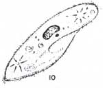
Protozoa as a predatorial parasite

Monogenea are large protozoans such as "Ich", Epistylis, Chilodonella, and copepod larvae, and can be seen in as little as 10x magnification on a standard microscope.

Epistylis
Epistylis
From Epistylididae, Epistylis with additional images and classification notes.

Fish inflicted with Epistylis infection
From NC Wildlife, Common Fish Diseases
"There are many factors that can cause a fish to get sick or develop sores. Fish are constantly exposed to various bacteria, fungi, viruses and parasites that occur naturally in the water. Generally the fish can deal with these with little or no problem. However, if fish are stressed or weakened by poor environmental conditions, they may not be able to fight off disease and may become sick. Some common diseases encountered in wild fish in North Carolina include white fuzzy patches on the skin caused by fungi or protozoans, red sores caused by bacteria, and black or yellow spots in the flesh of certain fish species caused by parasites. Some causes of stress include traumatic injury, spawning activity, rough handling and changes in water quality. When water temperature increases and/or fish are spawning, they are more likely to develop signs of disease. In most cases, the fish recover when conditions improve and stressors are reduced. Disease outbreaks typically don’t have a serious population level effect on natural fish populations. Usually only a small portion of a population will be seriously affected by a disease, and the population will rebound quickly."

Chilodonella
Chilodonella
Even one parasite warrants immediate action
The parasite Chilodonella is a major threat to fish health and finding even one parasite warrants immediate treatment. Chilodonella is a potentially dangerous parasite for two reasons. First, unlike many parasites, it has a wide range of temperature tolerance and outbreaks often occur at low temperatures when fish are least able to resist. Secondly, despite is relative small size it, is potentially more dangerous than Ich, because in the initial stages there are no readily visible signs of its presence.
From Chilodonella, A dangerous parasite

Chilodonella
Discus infested with the parasite Chilodonella, from Handbook of Fish Diseases
by Untergasser and Axelrod
"Chilodonella is a single cell microscopic parasite that attacks a fish skin and gills. The fish will rub against objects and become inactive. If the gills are affected they will stay near the water surface and gasp for air. Cloudy spots develop on the skin. The skin patches turn white and begin to disintegrate (usually over a couple day period). This opens the door to secondary infections and/or fungus. This parasite can swim and can attack other fish in the same aquarium. In aquariums with more fish, it is likely to spread rapidly. Poor water conditions also accelerate this disease. It can be introduced with live foods or from ponds or even on a plant."
From Chilodonella Parasite

In aquaculture, understanding larval stages and life cycles of parasites is necessary. To verify a diagnosis, parasites can be killed and preserved in 10% formalin and sent to a specialist.

Ichthyophthirius multifiliis
Life cycle of Ichthyophthirius multifiliis, from University of Florida
Ichthyophthirius multifiliis ("Ich" known as white spot disease) is a ciliate characterized by its relatively large size, in comparison with other protozoans.

Ichthyophthirius multifiliis
Cause of "ick" in fish. Large macronucleus can be seen in histopath sections of fish tissues.
From Clinical Parasitology, Oklahoma State University

Ichthyophthirius multifiliis is a devastating parasite which affects channel catfish and sometimes destroys entire populations. It is not host specific and may affect a variety of cultured finfish species. It burrows under the skin of fish, causing white specks that can sometimes be seen with the unaided eye.

Ich
Image modification of Ichthyophthirius multifiliis and is characterized by its relatively large size and horseshoe-shaped nucleus (Fundamentals of Aquaculture, Avault)

After maturing, the adult parasite called a trophozoite, leaves the fish and becomes free swimming for up to 6 hours, eventually attaching to substrate.

Trophozoite
Icthyophthirius multifiliis trophozoite from freshwater fish. Section through the surface of this ciliate, revealing the pocketed surface from which the cilia emerge. A number of basal bodies can be seen as well as numerous secretory bodies. 35,000x
From workforce.cup.edu

A membrane is secreted over the organism and cyst undergoes multiple fissions. As many as 1000 or more young, called tomites are produced. At temperature of 77 F, development into mature tomites can be completed within as little as 12 hours. When the cyst ruptures, tomites will begin the process of seeking a host, then penetrate the tissue of the fish host by cilary action and aid of an enzyme. Once penetrated, tomites mature into trophozoites and feed on the cell-tissue and fluid.

Ichthyophthirius multifiliis
Infected with Ick
"A common sign of beginning stages of Ich infection are what is called "flashing", where fish will swipe against aquarium decorations or the gravel at the bottom of the tank in an attempt to seemingly scratch themselves. Only one or two small colonies will appear at first, and be very difficult to identify until this infection advances to near maturity." (fishtankguy.com)
More informaiton is available on Icthyophthirius multifiliis at an earlier entry on Hybrid Striped Bass Culture.
Trichodina spp. is a ciliate which occurs among virtually all species of cold and warmwater fish, and includes both fresh and saltwater fish.

Trichodina
Trichodina, a ciliate and parasite
Image from source in Russian language, on Parasites.

This parasite may cause extensive mortalities especially in fingerlings and is sometimes associated with poor water quality and accumulation of waste. Epizootics (meaning epidemics in animals, root origin from the Greek, "epizoon" which refers to one animal living on the surface of another) may occur throughout the year though more common in spring and fall.

Trichodina
"Trichodina is a surface symbiont/parasite of fish. It has a broad oral disk surrounded by membranelles and an adhesive base." From Introduction to Ciliata

Fish who are infected may begin showing white to whitish-gray blotches and copious amounts of mucus may be present, along with milky and opaque tail fins. Infected fish may congregate near incoming water and become lathargic. Heavy parasitic infestation can result in excessive mucus production which impairs respiration and results in possible suffocation, though sufficient levels of dissolved oxygen may be present in the water. Related genera affecting fish include Trichodinella and Tripartiella.

Ambiphrya
Ambiphrya
From Fish Parasite Images, images from the collection of Dr. Thomas L. Wellborn, Jr.

Ambiphrya (Scyphidia) and Apiosoma (Glossatella) are similar parasites. They are associated with high concentrations of organic matter and found attached to plants, rocks, and other substrate. They are found on all cultured warmwater fish and occasionally on salmonids, and are mostly a problem with fry and fingerlings. Infected fish may not eat and may swim lethargically near the surface. Because of hemorrhages on the gills (including possible hermorrhaging of the body) Ambiphrya causes increased sensitivity to low levels of dissolved oxygen.

Epistylis, is a stalked protozoan and has a row of cilia at the apical end used for drawing food into the main body. Under microscope the stalks contract periodically which aid in proper identification. In the western United States, Rainbow trout are common host to the Epistylis parasite, and may occasionally occur on channel catfish, and will affect other finfish species. Common symptoms in scaled fishes are erosion of skin, scales, and spines results in bloody lesions, hence the name "red sore" disease. Other symptoms include hemmorrhaging and excessive localized mucus production. Infestations in channel catfish involve spines and bones that underlie the skin on the pectoral girdle, head, and fins. Epistylis is associated with a high concentration of pollution and/or organic matter. Infection will cause infested fish to flash. Infected fish eggs may appear "fuzzy".

Chilodonella
Chilodonella from Fish Parasite Images

Chilodonella is found on the gills and skin of finfish and distinct in parallel rows of cilia along the body margin. Chilodonella is a ciliate with a round to heart-shaped, depressed body. It is colorless and flattened, creeping rapidly across the fins, gills, and body of its host. Chilodonella is typically a coolwater parasite although one species, C. hexatichus, causes mortality at water temperatures up to 70°F.

Ichthyobodo
Ichthyobodo from Fish Parasite Images

Ichthyobodo is among the smaller protozoan parasites and often overlooked because it can only be detected under high magnification. Characteristically it is tear-drop shaped and has a "fluttering" appearance, like a tree leaf in a breeze when on gills or skin of the host fish. Ichthyobodo has one pair of flagella attached to the small rounded blepharoplast (small mass of chromatin embedded in cytoplasm at base of flagellum), but two smaller flagella appear before division.
Two species of Ichthyobodo have been documented, I. necatrix and I. pyriformis. I. necatrix is the most common of the two, is widely distributed throughout the United States and is common among young trout and salmon, where it is one of the most destructive ectoparasitic (parasites that live on the exterior of another organism) protozoans. Channel catfish fry and fingerlings are especially susceptible to this parasite.

Ichthyobodo necatrix
Ichthyobodo necatrix from Parasitic protozoa found on fish gills
"Ichthyobodo necator (Costia necatrix) is often found on the gills of young juveniles in the hatcheries in cases of inefficient incoming water treatment (filtration)."

I. necatrix attaches to the host body using a flat disc, extending into the host cell. Portions of the host cell are engulfed and brought into the parasite as food vacuoles ("One type of vacuole is the food vacuole, which is a temporary vacuole containing food that is obtained through phagocytosis ["cell eating"]. In addition, lysosomes recognize these food vacuoles and fuse with them for digestion of food particles. Without food vacuoles, a cell would not be able to be sufficiently nourished." From Vacuoles).
Often called the blue-slime disease, I. necatrix causes a grayish-white to bluish film to form on the skin of its prey, which comes from excess mucus production.

I. pyriformis infests gills and body and is found primarily on trout. It is pear-shaped and moves in rapid spiral pattern that is differentiated from darting movements of I. necatrix.

Dinoflagellates are identified by a groove around the middle of the cell with a flagellum lying in the groove. Oodinium sp., a well-known dinoflagellate found in fresh, brackish, and saltwater fish. Disease caused by Oodinium is often referred to as "velvet disease" since the parasite may become so abundant that a fine yellowish sheen appears.

Oodinium
Oodinium from Freshwater Disease and Treatment Chart

This parasite attacks the host' gills and skin and may be found attached to intestinal mucosa. The adult attaches to tissue with root-like appendages. Non-motile adults have a yellowish hue and frequently form clusters. Upon reaching maturity, they drop off and attaching themselves to a hard surface in the substrate, and begin multiplying, forming motile dinospores. Once a new host is found, their flagella disappear, and anchoring themselves to the host tissue with root-like appendages, until reaching maturity. Fish which are infested may scrape themselves showing signs of suffocation. A flashlight in a dark room may be used to inspect fish to check and see if light reflects from the Oodinium parasites.

Glenodinium
Glenodinium from Great Lakes Water Life Photo Gallery, Algae Dinoflagellates

Glenodinium, a dinoflagellate, has been associated with mortalities in channel catfish. It is normally a free-living algal cell which may develop into heavy bloom and give water a brown hue. Cells of Glenodinium may become entrapped in lamellae of fish gills causing a proliferation of tissue.

Trichophyra
Trichophyra
"Trichophyra piscium belongs to a Phylum of protozoan organisms all of whom possess cilia (‘hairs”) during at least one stage of their lifecycle. Most species are free-living aquatic organisms that feed on bacteria, some are commensal organisms, living on but not harming the host, and cleaning bacteria off the host’s body. Several species of Trichophyra have been identified as commensal organisms living on the gills of freshwater fishes. Large numbers of Trichophyra may cause gill irritation, or may cause the fish to secrete excessive gill mucous decreasing it’s respiratory and osmoregulatory efficiency. Some Trichophyra have been found parasitizing blood from the fish’s gills. In April 2002, 14-month old lake trout (S. namaycush) at the Maine Department of Inland Fisheries and Wildlife Enfield fish hatchery were acting as though they had gill parasites. Fish with gill parasites will rub their heads, and bodies against hard surfaces presumably to dislodge the organisms. Upon examination of the affected fish, Trichophyra piscium was identified in large numbers infesting the fish’s gills. The fish were treated as directed by MDIF&W’s fish veterinarian, and the problem was resolved."
From Protozoa: Trichophyra piscium

Trichophyra is a suctorian parasite infests gills of warmwater fish, and is distinguished by its round body and suctorial tentacles during the adult stage. It feeds on both passing protists as well as epithelial cells ('Epithelial' tissue works as a covering and lines organs in the body.) Gills may swell and become eroded, and anemia may follow. Affected fish become lethargic, may stop feeding, and may gather around inflowing water. Trichophyra populations may reach large numbers in an environment which contains high organic levels and low temperature.

Sporozoan protozoans are responsible for serious disease in fish. This class of protozoans is known by the spore morphology and by the number and location of polar capsules containing coiled filaments. Sporozoan parasites distributed in two basic groups, myxosporidians with two or more polar capsules and microsporidians with one polar capsule. Pathologists also recognize two other groups, coccidia and haemosporidea. Schäperclaus (1991), however pointed out that classification of sporozoa is difficult and that members of sporozoa have a single common feature, the formation of spores. Beyond that, classification is open to conjecture. Sporozoans tend to be host and tissue specific, having complex life cycles, and are untreatable. Life cycles of both myxosporidians and microsporidians begins at the spore level. When the host dies the spore drops to the bottom or accidentally eaten. Its polar filament(s) is used to attach to the gut wall. In miscosporidia, DNA nuclear material enters host cells through the everted polar filament. The parasite may eventually transfer to the definitive loci, or it may be carried there by the white blood cells. Once at the loci, sporozoan parasites (now termed trophozoites) divides (shizogony) and fuse (sprorogony), forming a mass of spores responsible for disease. (Fundamentals of Aquaculture, Avault)

Myxosoma cerebralis, a myxosporidian, causes "whirling disease" among salmonids, (diseased fish swim in circles). The parasite enters through any external opening, damaging the cartilage in the axial skeleton of young fish which intereferes with the function of neural structures and coordination. When fish are affected, they whirl as if chasing their tails. At one time whirling disease was the cause of catastophic losses in trout culture in central and north Europe, however today it is no longer considered a serious threat due to measures to control it. This disease has occurred in certain eastern states and Nevada.

Henneguya xenoma
Chain pickerel gill arch with Henneguya xenoma
When a xenoma ruptures, millions of Henneguya spores are released. The spores drift in the water and attach to a new host with a grappling hook-like organ called a polar filament. Once attached to a new host, the organism forms a new xenoma and begins to multiply. Fish veterinarians, culturists, biologists and others concerned with fish health may treat infected fish with chemotherapeutic agents or surgical removal. Unfortunately, many parasites, including Henneguya, are not easily controlled by any therapeutic procedure, thus prevention remains the best medicine. Henneguya is a fish parasite that seldom causes severe harm to the host. External examination may reveal cysts in the skin and gills; whereas, internal lesions may be found on the liver, heart, kidney, spleen or any other organ. Infections are usually not life threatening to the fish unless they impair the function of a vital organ. (From Myxosporidiosis: Henneguya sp. infestation)

Henneguya, found on freshwater finfish, is a myxosporidian having two polar capsules and a long-tail extension of each spore shell.

Henneguya
"Microscopic examination of the Henneguya sp. “xenomas” reveal tadpole shaped unicellular organisms with two eye-like polar capsules inside."
(From Myxosporidiosis: Henneguya sp. infestation)

Several apparent site-specific forms of Henneguya have been noted on channel catfish, on the skin and three on gill filaments. On the skin, a papillomatous form creates large lesions on body and fins, and nearly half the body may be affected. The second form creates a pustule or blister. Both forms may disfigure fish, but dressed fish show no signs of the parasite. The third form is of minor importance, occuring as a white cyst on the adipose fin of channel catfish fingerlings. On gills, an interlamellar form may cause extensive damage to channel catfish fingerlings. The second form usually has a few intralamellar cysts per filament, but does not pose a major problem. The third form produces discrete visible cysts on the gills. Henneguya has also been found in channel catfish viscera. (Fundamentals of Aquaculture, Avault).

Protozoan parasites and commensals of freshwater crawfish, prawns, and shrimp may be grouped as gregarines, microsporidians, ectocommensals, body invaders, and asptome ciliates. (Fundamentals of Aquaculture, Avault)

Gregarine protozoans, such as Nematopsis sp. have been observed in digestive tracts of shrimp in either the form of trophozoite or gametocyst. Life cycles of this parasite involve marine snails and/or clams. Trophozoites attach to the intestinal wall to asorb food, though over-all harm to the host is generally minor. Penaeid shrimp with Microspordian infestation can suffer from a chronic disease known as "cotton" or "milk" shrimp. Infestation is found throughout the musculature, depending on the species of microsporidia, or may be observed in particular tissue and organs. Microsporidians are found present in spore form with an envelope enclosing the spores in some species such as Pleistophora and Thelohania, but not all do. Nosema is one particular genus that lacks such an enclosing envelope. Infected shrimp may be active and feed normally, however microsporidians are suspect of inhibiting egg production in shrimp. Both macrobrachium and crawfish are susceptible hosts to microsporidians, though generally it is not considered a major problem in the United States. Microsporidian infection of crawfish in Europe is known as "porcelain disease". Spores are found spread throughout the musculature in heavily diseased crawfish and prawns. Research in areas of life cycles of microsporidians and their relation to crustaceans is incomplete.

Ectocommensals
"Protozoans such as Vorticella, Zoothamnium, Epistylis, Acineta and Ephelota. Affected shrimps are restless and their locomotion and respiratory functions are hampered. In heavily infected juvenile and adult shrimps, one can observe fuzzy mat-like appearance due to ciliate fouling. Maintain good water quality. Reduce organic load and silt in water exchange with good quality water."
From Protozoan Fouling, Indian Ocean.org

Ectocommensals are comprised of a variety of protozoan species which live on and/or attach to the surface of the body and the gills of their host. Common parasitic genera associated with crawfish and prawns include Epistylis, Zoothamnium, Lagenophrys, Corthunia and Acineta. Less common genera are Vorticella, Vaginicola and Opercularia.

Zoothamnium
Zoothamnium
"Zoothamnium niveum colonies on wood debris in the Indian River Lagoon. Zoothamnium niveum is a colonial Protist that reaches 2 - 3 mm in height. Individual zooids have an inverted bell shape and measure approximately 120um in height. The contractile vacuole is located below the peristomial lip. From Zoothamnium niveum, Smithsonian Institute.
"Zoothamnium prefers the gills (Johnson, 1978). Only rare incidences of heavy fouling affected the prawns adversely. Prawns have an increased oxygen demand just prior to moulting and heavy fouling can be associated with mortality due to anoxia. (Fisher, 1977)." (Fundamentals of Aquaculture, Avault)


Lagenophrys
Lagenophrys
"Lagenophrys limnoriae attached to the pleopods of Limnoria quadripunctata from the UK." From Institute of Marine Sciences, University of Portsmouth


Corthunia
Corthunia
"Hall (1979) found that Corthunia sp, Epistylis sp. and Vorticella sp. were the most common peritrichous ciliates in cultured prawns. Common sites of infestation are the body, eye stalk, antenna, uropods and egg masses. Thelohania, a microsporidian, has been reported in various species of marine shrimps but rarely in freshwater prawns. Areerat (1988) reported one case of microsporidia infection in the opaque muscular tissue of Macrobrachium."
From Diseases of the Freshwater Prawn, Aquatic Animal Health Research Institute

Freshwater Corthunia sp. is found on crawfish and occasionally prawns.


Acineta
Acineta
"The Suctorian Acineta: feeding."
From Micrographia
Acineta sp. is commonly found in brackish water on prawns.

Epistylis, Zoothamnium and Lagenophrys are found in both fresh and brackish water on crawfish and prawns. Lagenophrys is more prevalent on prawns than crawfish.
Body invaders are protozoans that on occasion are discovered wandering about the body of their host, but if this is a case of "accidental parasitism", and whether the crustacean serves as a paratenic host, is still unclear.
Apostome ciliates (considered to be commensal) are commonly found on crawfish and prawns and have been noted infrequently in the resting stage of shrimp. Apostomes will encyst on crustaceans' exoskeleton. When crustacean molt, protozoa hatch and feed on fluids released on the shed exoskeleton. They give the appearance of tiny transparent bubbles. Following reproduction, apostomes begin the search for a host. Apostome genera commonly found in North America are Hyalophysa, Gymnodinioides and Terebrospira.

In the North American oyster culture, several serious protozoan diseases have been identified. Haplosporidium nelsoni, known as MSX before 1966, is the cause of "Delaware Bay disease" in the American oyster and likely affects other species. Oysters which are infected become emaciated and have weak shell enclosure and recession. This disease is particularly lethal for oysters, and influenced by high salinity. During dry years with high salinity (mid 60's), H. nelsoni caused high mortalities to oyster beds in Maryland, and rainy years (71-74) the disease remained confined to Mobjack Bay and the lower Chesapeake Bay. A salinity of 15 ppt or higher puts oysters at risk for the disease which may remain hidden up to 9 months. Some outbreaks encompassed mortalities up to as high as 50%-60%.
Haplosporidium costale, causes seaside disease which occurs in high salinity waters of over 25 ppt. Oysters affected by this disease characteristically fail to add new "bill" or shell, resulting in overall poor health. This disease occurs in Long Island Sound to Cape Charles and sometimes the Delaware and Chesapeake bays. H. costale has no tolerance for salinities below 25 ppt. Juveniles under two or three years may survive an outbreak, but killing up to as many as 20-50% of mature oysters. This parasite has been under study for more than thirty years because of its seriousness.
Oyster Diseases - Comparisons of Poor Health
Oyster DiseaseOyster Disease
Perkinsus marinus known as "Dermo" is common to the Gulf of Mexico and lower Chesapeake Bay and Haplosporidium nelsoni, known as MSX before 1966, is the cause of "Delaware Bay disease" in the American oyster and likely affects other species.
Oyster DiseaseOyster Disease
Images from Dermo and Oyster Research and Restoration.

The parasite invades the gut epithelium, possibly through the mantle. Once the epithelium is destroyed, the parasite is further distributed by blood throughout the remainder of the oyster. It inhibits normal gonadal development and severely emaciates affected oysters. Infections typically rise during warmer months and decline during colder months and salinities below 15 ppt, infections and resulting mortalities are thus reduced. Protozoans Marteilia refringens and Bonamia ostreae have contributed to serious problems in European waters with the oyster, Ostrea edulis.
Haplosporidian protozoans have been reported for moribund mussels in North America, but presently longterm implications remain unclear. Appearing to be an isolated event, extensive mortality of mussels on Prince Edward Island occurred because of infestation by a haplosporidian protozoan, identified as Labyrinthomyxa sp.
With increasing aquaculture of mussels (primarily Mytilus edulis), clams (various species), and scallops (several species), protozoan diseases are attracting more attention.

Reference Works Consulted and Cited
1. Fundamentals of Aquaculture, Avault
2. Ecology and Classification of North American Freshwater Invertebrates, Thorp and Covich
3. Zooplankton of the Atlantic and Gulf Coasts, A Guide to Their Identification and Ecology, Johnson and Allen
4. Freshwater Microbiology, Grazing activities in the freshwater environment: the role of protozoa and invertebrates, David C. Siege
Read More »